Article Text

Statistics from Altmetric.com
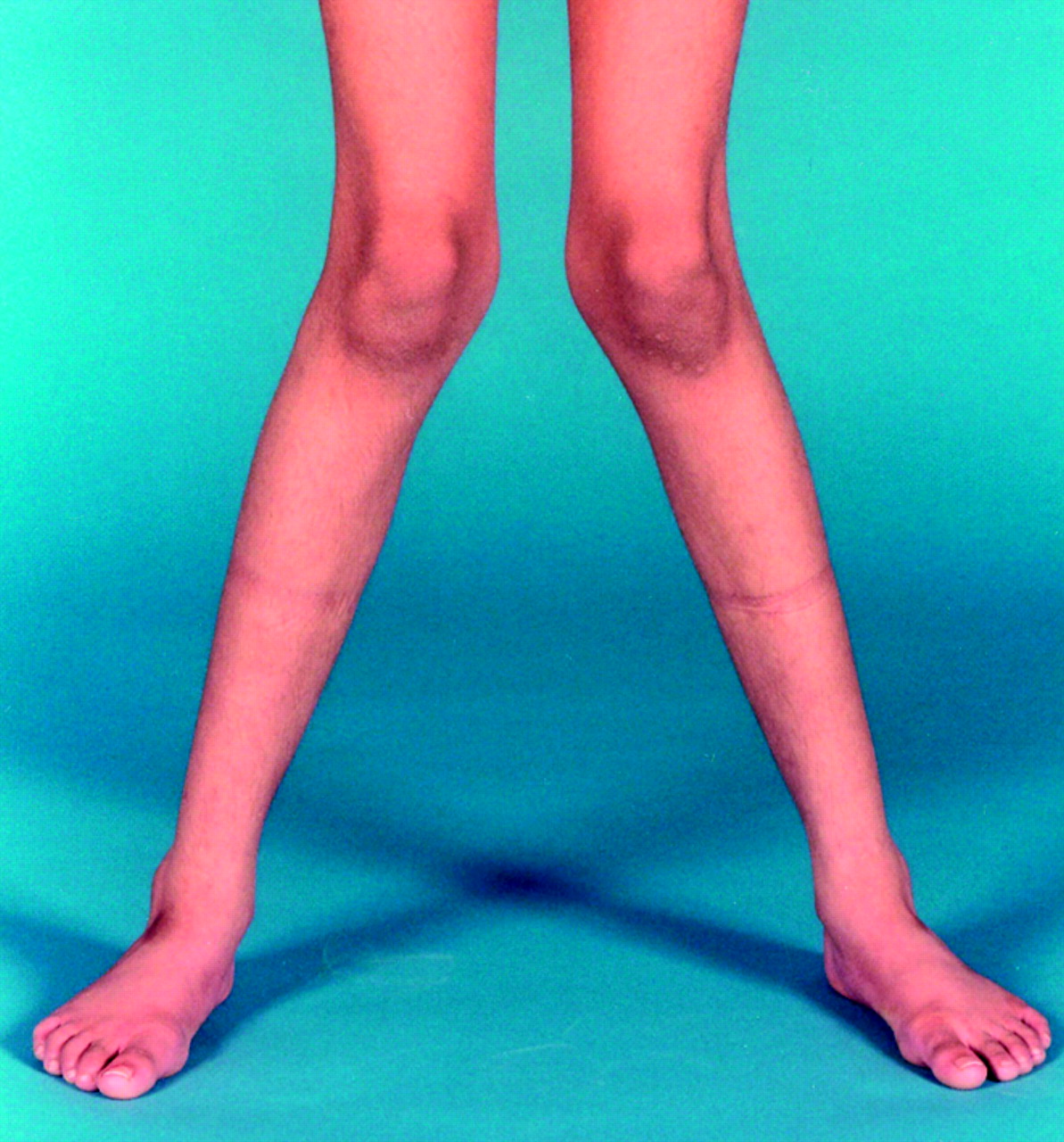
We have previously drawn attention to a resurgence of vitamin D deficiency rickets in young children of South Asian (Indian, Pakistani, and Bangladeshi) and Middle Eastern origin, living in the UK.1,2 We now describe nine (one male) non-Caucasian, adolescents (age 11–17 years) who presented with symptomatic vitamin D deficiency between 1997 and 2002. Table 1 summarises their ages, ethnic origins, clinical symptoms, signs, and relevant biochemical findings at presentation. All presented with symptoms of vitamin D deficiency, which included lower limb pains, difficulty in walking or climbing stairs, carpopedal spasms, and hypocalcaemic convulsions. Clinical signs included positive Chvostek sign, inability to stand up unaided from a squatting position due to proximal myopathy, bowed legs (genu varum), and knock-knees (genu valgum). Three patients (cases 1, 6, 9) had radiological changes of vitamin D deficiency with widening and fraying of metaphyses, but these changes were not as severe as those seen in toddlers with vitamin D deficiency rickets. As shown in table 1, biochemical features of the disease included low serum concentrations of 25-hydroxycholecalciferol (25(OH)D; a measure of an individual’s vitamin D status), increase of serum alkaline phosphatase activity for the age (except in cases 2, 5, and 7), secondary hyperparathyroidism leading to hypophosphataemia (except in cases 7 and 8), and formation of normal, or supranormal serum concentrations of 1,25 dihydroxycholecalciferol (except in case 5). The presenting symptoms in all patients resolved after treatment with oral vitamin D. However, unlike in younger children, genu valgum or genu varum (fig) do not fully correct after treatment with vitamin D after the long bone epiphyses have closed; the residual deformities require surgical correction.
Vitamin D is necessary for adequate bone mineralisation and its deficiency results in rickets in children and osteomalacia in older adolescents and adults. In humans, the main source of vitamin D is that produced by the action of solar ultraviolet light B radiation acting on 7-dehydrocholesterol in skin. Small amounts are also derived from dietary sources: oily fish, eggs, and fortified foods, such as margarine and breakfast cereals. The Committee on Medical Aspects of Food Policy (COMA)3 recommends that all pregnant women, lactating mothers, and children up to the age of 5 years who are at risk of vitamin D deficiency should be encouraged to take supplements. However, there are no recommendations for vitamin D supplementation in adolescents, although the COMA does recommend provision of vitamin D supplements to vulnerable older children.
Adolescence is a critical period of skeletal mineralisation, when over 35% of the peak bone mass (PBM) of a mature adult is accrued during the four years surrounding the peak pubertal growth spurt.4 It is widely accepted that subjects who attain a lower PBM at maturity have a higher risk of sustaining osteoporotic fractures in later life.5 Thus, inadequate skeletal mineralisation secondary to vitamin D deficiency during this period might compromise the acquisition of PBM and thus potentially increase the risk of osteoporosis in later life. We therefore recommend that a prospective study is needed to determine the prevalence of vitamin D deficiency among adolescents whose exposure to sunshine is limited for cultural and religious reasons.
Demographic and clinical data of the patients
Severe knock-knee (genu varum) deformity in case 8.