Article Text

Abstract
Aim of the study Tissue engineering appears as a potential alternative treatment for long-gap esophageal atresia, for which otherwise there is few treatment options. However, lack of biocompatibility of synthetic materials used in preclinical experiments hampered regeneration of functional muscle layers, eventually leading stricture in the oesophagus or displacement of the material. To overcome this problem, we took advantage of cell-based bioengineering in which progenitor cells with myogenic potential were seeded into extracellular matrix (ECM) scaffolds obtained by decellularization of piglets’ oesophagi.
Methods Oesophagi were harvested from piglets and decellularized through two cycle of the detergent-enzymatic treatment (DET) in tailored bioreactors. Mesoangioblasts (MABs) and fibroblasts (FBs) were isolated from the biopsy of human skeletal muscle and expanded. The cells were co-seeded into the ECM scaffolds, at a proportion of 85% MABs and 15% FBs. The seeded oesophagi were cultured in the dynamic condition, in which custom designed bioreactors generate continuous flow of cell culture media inside the lumen. Incubation was maintained for up to 10 days.
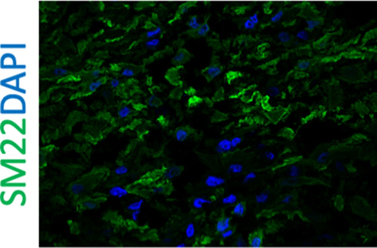
Results Histology of decellularized oesophagi showed absence of the native cells. The ECM construction of the scaffold was comparable to native oesophagus, preserving distinctive layers. Immunohistochemistry of the repopulated oesophagi identified migration of the cells from the injected sites and differentiation of MABs to smooth muscle cells with expression of SM22 and αSMA. Co-seeded FB distributed along the scaffold maintaining the initial proportion of cells (figure 1).

Masson trichrome staining of recellularized oesophagus
Immunostaining of recellularized oesophagus
Conclusions We demonstrated efficient decellularization by DET and migration and differentiation of MABs in the ECM scaffold promoted by co-seeding with FBs. Utilization of an oesophagus scaffold from piglets, which has a similar size to human neonates’ oesophagus, will be promising strategy for clinical application of regenerative medicine in humans.