Article Text

Abstract
Background—In several diseases there is a relation between deficiency of neutrophil granulocytes and granulomatous lesions. Recently, in glycogen storage disease type Ib, this relation has been supported by the beneficial effect of treatment of enteritis with granulocyte-macrophage colony stimulating factor.
Aim—To investigate whether chronic granulomatous disease could be treated according to the same principle.
Patients and methods—Inflammatory lesions were monitored in two brothers with chronic granulomatous disease demonstrated by very low superoxide production in neutrophil granulocytes. The two patients were treated with recombinant human granulocyte colony stimulating factor on three occasions when the disease was active.
Results—In one patient, remission of an inflamed stenosis of the colon sigmoideum was shown by granulocyte scintigraphy after one month of treatment with granulocyte colony stimulating factor. In the other patient, remission of colon disease and later of a non-malignant tumour in the right lung hilum was shown by colonoscopy and computed tomography scans respectively.
Conclusion—Remission of inflammatory lesions in two brothers with chronic granulomatous disease was induced by granulocyte colony stimulating factor on three occasions. The mechanism for this effect is not known. The result is similar to the response found in patients with leucocyte deficiency due to glycogen storage disease type Ib.
- chronic granulomatous disease
- enteritis
- granulocyte colony stimulating factor
Statistics from Altmetric.com
Chronic granulomatous disease (CGD) is a group of inherited disorders characterised by defective superoxide production in circulating phagocytes leading to impaired bactericidal activity.1-5 The patients therefore suffer from frequent bacterial and fungal infections.1 The different clinical forms of CGD are related to genetic heterogeneity.6 A number of patients suffer from enteritis.7 ,8 Enteritis is also seen in other disease states with defective neutrophils.9-12 In glycogen storage disease type Ib,13-16 the association between the granulomatous lesion and neutrophil deficiency is supported by the recent demonstration that patients with enteritis benefit from treatment with granulocyte-macrophage colony stimulating factor.17 We report three incidents of clinical remission in two brothers with CGD during treatment with granulocyte colony stimulating factor (G-CSF). In parallel with the results from glycogen storage disease type Ib, this may serve as a model for the pathogenesis and new treatment strategies in other granulomatous diseases.
Case reports
METHODS
Recombinant human G-CSF was obtained from Roche, Denmark (Neupogen). Each treatment regimen consisted of subcutaneous injections of 0.3 mg recombinant human G-CSF daily for 30 days. Erythrocyte sedimentation rate and orosomucoid and leucocyte counts were recorded once a week during the treatment periods. The oxidative metabolism of neutrophils was assessed by phorbol myristate acetate stimulated nitroblue tetrazolium slide test, oxygen consumption, superoxide anion production, and chemiluminescence as previously described.18 Chemotaxis was measured in a Boyden chamber using N-formyl-methionyl-leucyl-phenylalanine (f-MLP) as chemoattractant.19 The effect of treatment on the leucocyte population distribution and membrane phenotype was assessed by differential counting and flow cytometric quantification of cluster of differentiation 14, 11b and 16 antigens on granulocytes and monocytes.
PATIENT 1
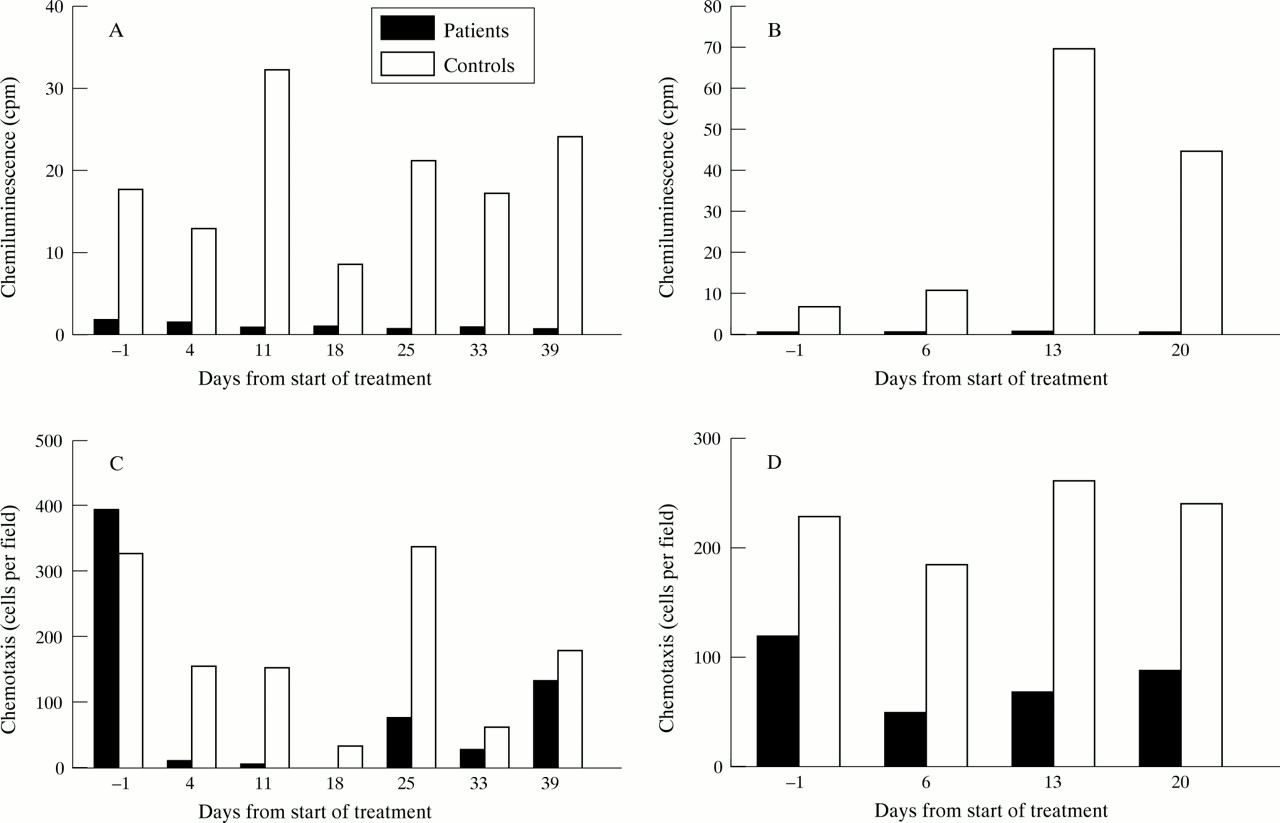
The patient had suffered from chronic inflammatory bowel disease since the age of 26. He reported having seven to ten stools per day, often containing blood. Barium contrast studies showed a stenosis in the rectum and inflammation of the sigmoid and descending colon. After exclusion of infectious causes, the patient was treated with prednisolone, salazopyrine, and azathioprine without clinical improvement. The symptoms did not change over the following three years. Repeated barium contrast studies gave unchanged results. CGD was suspected because of a similar diagnosis in the patient’s brother (patient 2). The diagnosis was confirmed by a weakly positive nitroblue tetrazolium test, very low oxidative metabolism of neutrophils and monocytes measured by chemiluminescence as well as low superoxide production in granulocytes after induction with f-MLP. Erythrocyte sedimentation rate and plasma orosomucoid were slightly elevated. The patient received G-CSF treatment for 30 days after three years of continuous clinically active disease. He reported a reduction in diarrhoea from ten to four stools per day and disappearance of bleeding. Colonoscopy and sigmoidoscopy were unsuccessful because of the severe sigmoidal stenosis at this stage. Instead, disease activity was monitored by granulocyte scintigraphy, which was performed before treatment, one month after initiation of treatment, and one, seven and 26 months after termination of treatment. Granulocyte scintigraphy was performed 24 hours after injection of 111In labelled granulocytes. Imaging was performed using a General Electric 600xR gamma camera (General Electric Medical Systems, Buc, France). Before treatment, clear inflammatory activity located in the colon sigmoideum was observed on the granulocyte scintigram, which was almost normalised after one month of treatment. No scintigraphic relapse was seen one, seven and 26 months after termination of treatment. Erythrocyte sedimentation rate and plasma orosomucoid did not change during treatment. The treatment induced neutrocytosis (mean 25 (range 15–27) × 109 per litre). Oxidative metabolism measured as f-MLP induced chemiluminescence remained low (fig 1A). f-MLP induced chemotaxis showed a decrease on initiation of treatment, followed by a slow increase during the course of treatment; however, normal values were not reached (fig 1C). The cluster of differentiation 14 antigen increased from 1250 to 25 000 molecules per neutrophil, indicating a shift in neutrophil mobilisation and activation of the bone marrow pool (fig 2).

N-Formyl-methionyl-leucyl-phenylalanine (f-MLP) induced chemiluminescence in patient 1 (A) and 2 (B) during treatment with granulocyte colony stimulating factor (days 1–30) in comparison with samples from healthy individuals analysed on the same day. f-MLP induced chemotaxis in patient 1 (C) and 2 (D) during treatment with granulocyte colony-stimulating factor (day 1–30) in comparison with samples from healthy individuals analysed on the same day.

Numbers of cluster of differentiation 14 antigens on neutrophils in patient 1 during treatment with granulocyte colony stimulating factor (days 1–30).
PATIENT 2
The patient was treated with G-CSF on two occasions when disease was active. At the age of 24 years the patient had recurrent dermal abscesses, and a granulomatous lesion of a lymph node at the right lung hilus was found. He was treated for suspected tuberculosis, and regression of the lesion occurred during antituberculosis treatment. Three years later, CGD was suspected on the basis of clinical presentation, and the diagnosis was confirmed by the same methods as used for patient 1. The patient was admitted to our department at the age of 28 because of abdominal pain and meteorism. Colonoscopy and barium contrast studies showed an ulcerated tumour of the left colonic flexure. Biopsy specimens showed chronic non-specific inflammation. Erythrocyte sedimentation rate and plasma orosomucoid were elevated. The patient was treated with G-CSF for 30 days. During the treatment period, he reported relief of abdominal symptoms. A colonoscopy two weeks after initiation of treatment showed improvement in the ulcerative inflammatory process of the left colonic flexure with complete mucosal healing at four weeks. The patient was asymptomatic until one year later, when he was readmitted with cough, expectoration, and haemoptysis. Radiographs showed prominence of the right lung hilus, and computed tomography scans suggested a lung tumour at this site. Bronchoscopy showed no sign of malignant disease, and biopsy samples from the tumour area at mediastinoscopy were without malignancy. Three months later a computed tomography scan was repeated with no detectable change. The patient was again treated with G-CSF for 30 days. The symptoms of cough and haemoptysis disappeared two weeks after initiation of G-CSF treatment. Computer tomography demonstrated a clear regression of the process at the right lung hilum. The size of the granulomatous lesion decreased from 4.5 × 2.5 × 2 cm to 1 × 2 × 1 cm. Eight months later it was undetectable. Erythrocyte sedimentation rate and plasma orosomucoid were unchanged during the first treatment period, decreased during the second treatment period, but did not normalise. The treatment induced neutrocytosis (mean 24 (range 18–28) × 109 and mean 31 (range 28–36) × 109 per litre respectively). Oxidative metabolism measured as f-MLP induced chemiluminescence and superoxide production remained low (fig 1B). f-MLP induced chemotaxis showed a decrease on initiation of treatment, followed by a slow increase during the course of treatment (fig 1D). Cluster of differentiation antigens were not measured in this patient.
Discussion
On the basis of the very low but detectable stimulation of the oxidative metabolism by f-MLP, it was concluded that both patients suffered from the Xb− form of CGD.2 G-CSF has been shown both in vivo and in vitro to increase f-MLP induced chemiluminescence,20 ,21 but this seems not to be the case in CGD and therefore cannot explain the demonstrated clinical effect. A change in the properties of neutrophils did, however, occur since the number of cluster of differentiation 14 antigens increased substantially. Without activation, neutrophils only weakly express cluster of differentiation 14 antigens.22 Another possibility is that, although the single neutrophil remained partially deficient in respiratory burst activity, this could be compensated for by an increased number of cells as the result of stimulation of granulopoiesis. The finding of reduced chemotaxis during G-CSF treatment was expected.23 Our result is in parallel with reports of clinical remission of enteritis in glycogen storage disease type Ib by granulocyte-macrophage colony stimulating factor.17 These inflammatory processes seem to be modified by a mobilisation of inflammatory cells from the bone marrow. It thus appears likely that, in CGD and glycogen storage disease type Ib, inflammation induced by an antigen is not resolved.
In conclusion, remission of inflammatory lesions in two brothers with CGD was induced by G-CSF on three occasions. The mechanism for this effect is not known. The result is similar to the response found in patients with leucocyte deficiency due to glycogen storage disease type Ib.
Acknowledgments
Dr Hans Johnsen, Department of Haematology, Herlev Hospital, Copenhagen Denmark is thanked for analysis of surface antigens, and valuable comments on the manuscript.