Article Text

Abstract
Background: Gastrostomy feeding is a well established alternative method to long term nasogastric tube feeding. Many such patients have gastro-oesophageal reflux (GOR) and require a fundoplication. A transgastric jejunal tube is an alternative when antireflux surgery fails, or is hazardous or inappropriate.
Aims: To review experience of gastrojejunal (G-J) feeding over six years in two regional centres in the UK.
Methods: Retrospective review of all children who underwent insertion of a G-J feeding tube.
Results: There were 18 children, 12 of whom were neurologically impaired. G-J tubes were inserted at a median age of 3.1 years (range 0.6–14.7) because of persistent symptoms after Nissen fundoplication (n = 8) or symptomatic GOR where fundoplication was inappropriate. Four underwent primary endoscopic insertion of the G-J tube; the remainder had the tube inserted via a previous gastrostomy track. Seventeen showed good weight gain. There was one insertion related complication. During a median follow up of 10 months (range 1–60), four experienced recurrent aspiration, bilious aspirates, and/or diarrhoea. There were 65 tube related complications in 14 patients, necessitating change of the tube at a median of 74 days. Jejunal tube migration was the commonest problem. Five died from complications of their underlying disease.
Conclusions: Although G-J feeding tubes were inserted safely and improved nutritional status, their use was associated with a high rate of morbidity. Surgical alternatives such as an Roux-en-Y jejunostomy may be preferable.
- percutaneous endoscopic gastrostomy
- gastrojejunal feeding tube
- G-J, gastrojejunal
- GOR, gastro-oesophageal reflux
- PEG, percutaneous endoscopic gastrostomy
- PEG-J tube, gastrojejunal tube inserted by percutaneous endoscopy
Statistics from Altmetric.com
- G-J, gastrojejunal
- GOR, gastro-oesophageal reflux
- PEG, percutaneous endoscopic gastrostomy
- PEG-J tube, gastrojejunal tube inserted by percutaneous endoscopy
Children with feeding difficulties frequently require supplementary or total enteral nutrition; this may be provided in a variety of ways.1 Percutaneous endoscopic gastrostomy (PEG) was first described in 1980 for long term enteral feeding in such children.2 A concomitant fundoplication is usually required in patients with severe symptomatic gastro-oesophageal reflux (GOR).3 For those in whom a fundoplication has failed, either because of recurrent GOR or gastric dysmotility, and where redo fundoplication is not appropriate, unlikely to be successful, or deemed too hazardous, an alternative approach to feeding is via a gastrojejunal (G-J) tube. Gastrojejunal feeding may also be advantageous in conditions such as acute pancreatitis,4 and in paediatric intensive care where gastroparesis may preclude nasogastric feeding but where small bowel function is preserved. There are only a few studies describing the use of G-J feeding tubes in children and there is little available information on outcome.5–7 We therefore reviewed experience of gastrojejunal feeding tubes in children at two regional paediatric centres.
MATERIALS AND METHODS
Case notes of all children undergoing insertion of a G-J tube at two regional paediatric units during a six year period (1994–99) were retrospectively reviewed. All children underwent prior assessment by a multidisciplinary team which included a paediatric gastroenterologist, specialist paediatric nurse, dietician, and paediatric surgeon. Demographic details, principal diagnosis, indications, technique of G-J tube insertion, and outcome data were recorded. The latter included complications associated with tube insertion, tube related problems, duration of G-J tube feeding, weight gain, and any subsequent surgical interventions. Children in whom surgical types of feeding jejunostomy were established were excluded from this analysis.
RESULTS
Between January 1994 and December 1999, 18 children (12 in Leeds and six in Manchester) had a G-J feeding tube inserted. The median age at G-J insertion was 3.1 years (range 0.6–14.7). Twelve children (66%) were neurologically impaired (table 1). Fourteen had undergone previous surgery: Nissen fundoplication in eight, PEG insertion in five, and an open gastrostomy in one. Four children had a G-J tube inserted using a percutaneous endoscopic technique (PEG-J)8; a G-J tube was placed through a pre-existing gastrostomy track in the remainder.5 The Freka gastrojejunal tube, which comprises an outer 15 Fr gastrostomy tube with a separate inner 9 Fr jejunal tube (Fresenius Bad Homburg, Germany) was used in nine patients; the 16 Fr integral MIC G-J tube (Medical Innovations Corporation, Milpitas, California) was inserted in the other nine (fig 1). Table 2 shows the indications for G-J tube insertion.
Diagnoses in children undergoing G-J tube insertion (n=18)
Indications for G-J tube insertion (n=18)

Fresenius component PEG-J tube (above) and the integral MIC PEG-J tube (below).
Outcome and nutritional progress
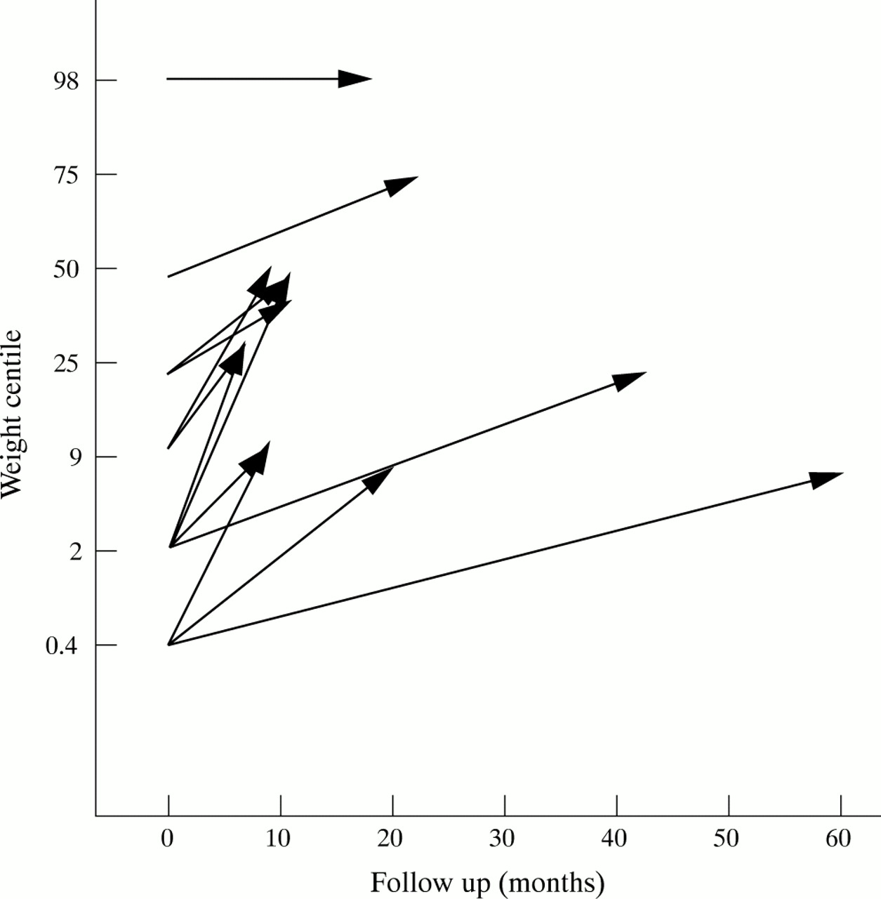
Five children died from complications related to their underlying condition between 6 and 18 months after G-J tube insertion: two from progressive underlying liver disease (one of whom had previously undergone liver transplantation); two from sepsis and multiple organ failure; and one from profound unexplained metabolic acidosis. All five showed good initial weight gain (from 3rd to 9th centile in three and 9th to 25th centile in two), but this was only temporary in the two children with liver disease. Of the 13 surviving children, 12 gained weight after insertion of a G-J tube (fig 2). At the end of the follow up period, five children continued to be nourished via their G-J tube, six were feeding satisfactorily via a PEG tube, one had undergone a Nissen fundoplication and gastrostomy, and one child had a Roux-en-Y jejunostomy.

Changes in weight centiles during G-J tube feeding (n=13).
Complications of G-J tube feeding
There were 65 tube related complications in 14 patients (table 3). One severely neurologically impaired child developed a pneumoperitoneum after tube insertion. No gastrointestinal leak was shown by contrast radiology or at subsequent laparotomy. She had persistent bilious gastric aspirates and retching and underwent a Nissen fundoplication and feeding gastrostomy two months later. She died six months later of irreversible metabolic acidosis of unknown aetiology. In one other child who had undergone multiple surgical procedures including liver transplantation, the tip of the G-J tube could not be advanced beyond the distal duodenum. Two children had intermittent diarrhoea and two others had recurrent episodes of aspiration; two of these also had intermittent, large, bilious gastric aspirates.
Complications of G-J tubes in 18 patients (65 complications in 14 children)
Mechanical tube problems were the most frequent complication; most of these necessitated reinsertion of the jejunal tube, either under general anaesthesia with endoscopic and/or fluoroscopic assistance, or after sedation in the radiology department. The number of tube changes per patient ranged from 0 to 12. The median interval between insertion and first tube replacement was 74 days (range 9–660). Jejunal tube migration was the commonest tube related complication, accounting for 75% of mechanical complications. There were eight episodes of complete inward migration of the tube. In seven of these, retrieval of the jejunal tube from the stomach was carried out endoscopically under general anaesthesia. In one case, a plain abdominal radiograph showed the tube in the caecum; the child was successfully managed expectantly.
DISCUSSION
Percutaneous endoscopic gastrostomy was initially described as a means of long term enteral feeding in children who were neurologically impaired and could not swallow or had inadequate calorie intake, or had severe musculoskeletal deformity posing a high anaesthetic and surgical risk from operations designed to achieve long term enteral access.2 As experience with PEGs increased, notable complications were identified, including symptomatic GOR in previously asymptomatic patients or worsening GOR necessitating an antireflux procedure.9–11 Although recent studies have suggested that the frequency of these problems may have been overestimated in the past,3,12 these complications nevertheless do occur with gastrostomy feeding and are more common in neurologically impaired children.9,11
In 1984, the PEG-J tube was introduced as a method of circumventing these problems.8 This was based on the assumption that placing the tip of the tube beyond the ligament of Treitz prevented duodenogastric reflux or GOR. G-J feeding tubes have since been used in children who are deemed too unfit for antireflux surgery or where fundoplication has failed.6,7,13 Such children include those with severe physical and neurological handicap who are at greater risk of surgical and anaesthetic complications and have an increased mortality from respiratory complications associated with gastrostomy tube feeding.14 In our series, G-J tubes were used in a highly selected group of patients. This is highlighted by the fact that the two senior surgical authors in Leeds (MDS and DCC) performed 108 Nissen fundoplications with gastrostomy, 184 PEGs, and 29 open jejunostomies in children with feeding difficulties during the six year study period.
Several techniques have been described for the insertion of G-J tubes in children.5–7 The G-J tube may be inserted as a single stage procedure by an endoscopist (a PEG-J tube) or a radiologist using a percutaneous technique. Alternatively, the G-J tube can be inserted through an existing gastrostomy track. In all cases the tube is manipulated into position using fluoroscopy and/or endoscopy. Difficulty intubating the jejunum is frequently encountered; this is usually attributed to unfavourable angulation of the gastrostomy track.5,15 The interventional radiological approach to insertion is usually performed under local anaesthesia with sedation.6,7 For the endoscopic technique, we recommend general anaesthesia for insertion of the gastrostomy tube, although jejunal tube insertion/reinsertion can safely be performed with the child awake, sedated, or under anaesthesia, depending on the anticipated complexity of the procedure. Irrespective of the technique, complications associated with insertion of the G-J tube have been uncommon, as in our small series of patients.7
G-J tube feeding in our patients had clear nutritional benefits. However, the use of G-J feeding tubes was associated with a high incidence of complications. There was presumptive evidence of duodenogastric reflux of bile in two children, and GOR in two others, despite correct positioning of the tube beyond the ligament of Treitz. The majority of complications were mechanical and included tube migration, fracture, dislodgement, and blockage. In adults, these problems have been reported to be much more frequent with PEG-J tubes than PEGs.16 Published experience of G-J tubes in children is limited.5–7 Chait et al reported their combined experience of percutaneously inserted gastrostomy and G-J tubes, but the complication rate of the latter, which accounted for just 14% of all cases, was not specifically stated.6 Albanese et al described their results after G-J tube insertion in 44 neurologically impaired children.7 Follow up data were available for 34 patients, 12% of whom experienced major complications (small bowel intussusception, persistent GOR, and pneumonia) and 44% minor complications. The latter were mostly related to jejunal tube migration which occurred despite a policy of elective change of the jejunal tube every three to four months. McHugh commented that the mean duration of jejunal catheter use was 54 days in a small group of children with pre-existing gastrostomies and feeding difficulties.5
Until these problems can be solved, for children who require long term artificial feeding and cannot be managed by a feeding gastrostomy with or without fundoplication, an open jejunostomy is preferable. Our initial experience with the Roux-en-Y feeding jejunostomy (Maydl's procedure) is encouraging. This technique appears to be a safe and reliable alternative to conventional jejunostomy techniques.17,18 Our current opinion is that the indications for G-J tube placement are few. Use of G-J tubes should be confined to children requiring short term jejunal feeding (who cannot be managed with a nasojejunal tube), and children with severe gastric dysmotility after fundoplication who cannot tolerate intragastric feeds because of intractable retching. Adequate weight gain can be achieved, but the inability to bolus feed necessitates continuous enteral nutrition for at least 14 hours per day, which is problematic for patients and carers.